

A recent study published on the bioRxiv* preprint server reports that vitamin D deficiency enhances disease severity, while adequate vitamin D supplementation reduces inflammation, after H1N1 and severe acute respiratory syndrome coronavirus (SARS-CoV-2) infections in mice.
Study: Vitamin D and the ability to produce 1,25(OH)2D are critical for protection from viral infection of the lungs. Image Credit: Festa / Shutterstock.com
Background
Low vitamin D status is associated with a poor prognosis in patients with acute respiratory diseases, such as influenza and the coronavirus disease 2019 (COVID-19). In fact, recent evidence has indicated that low circulating vitamin D (serum 25(OH)D, 25D) levels were associated with severe disease and higher mortality rates in COVID-19 patients. As a result, high-dose vitamin D supplementation has been recommended to reduce the severity of these infections.
The relationship between vitamin D levels and respiratory disease severity appears to be due to the antiviral activity of vitamin D that may be specific to each virus. For example, vitamin D, as well as its analogs, appear to induce the production of cathelicidin, which is an antimicrobial peptide that is released in response to viral infections. Conversely, vitamin D may also limit inflammatory responses by reducing interferon γ (IFN- γ), interleukin 6 (IL-6), and tumor necrosis factor α (TNF-α) levels.
Lung epithelial cells express the vitamin D receptor (VDR) and are regulated by 1,25D supplementation. Thus, vitamin D treatment has been shown to directly affect the lung epithelium and reduce the inflammatory response within the lungs.
Vitamin D supplementation and H1N1 infection
The researchers in the current study sought to further evaluate the lung response of vitamin D supplementation in treating both H1N1 and SARS-CoV-2 infection in vivo. For the H1N1 studies, the researchers utilized two different strains of mice, of which included wildtype (WT) and CYP27B1 knockout (Cyp KO) mice. Taken together, age and sex-matched mice were fed chow diets, which contained vitamin D (D+) or purified diets with (D+) and without (D-) vitamin D.
The CYP27B1 gene ensures the production of 1-alpha hydroxylase, which initiates the production of 1,25D, the active form of vitamin D. Because Cyp KO mice are unable to produce 1,25D, the researchers were able to assess the effects of both H1N1 and SARS-CoV-2 infection in the presence of a severe vitamin D deficiency, as well as how vitamin D supplementation might improve the inflammatory response in this environment.
Irrespective of the genotype, serum 25hydroxy vitamin D (25D) levels markedly varied amongst mice fed a D+ or D- diet. Nevertheless, 25D levels in D+Cyp KO mice were greater than those of D+WT mice.
Although all mice infected with H1N1 experienced respiratory distress seven days after infection, D+WT mice experienced the least amount of respiratory distress. All symptoms of respiratory distress in both WT and Cyp KO mice receiving vitamin D supplementation were resolved by day 10. Comparatively, both D-WT and D-Cyp KO continued to experience greater symptoms of respiratory distress that did not completely resolve by day 14.
In addition to symptom resolution, vitamin D supplementation provided the greatest benefit to WT mice in terms of their survival, as well as lung pathologies. Despite vitamin D supplementation in Cyp KO mice, their baseline level of damage within the lungs, combined with the respiratory effects of H1N1 infection, could not be recovered with vitamin D supplementation.
Taken together, vitamin D deficiency and the lack of 1,25D synthesis increased the vulnerability of mice to H1N1 infection.
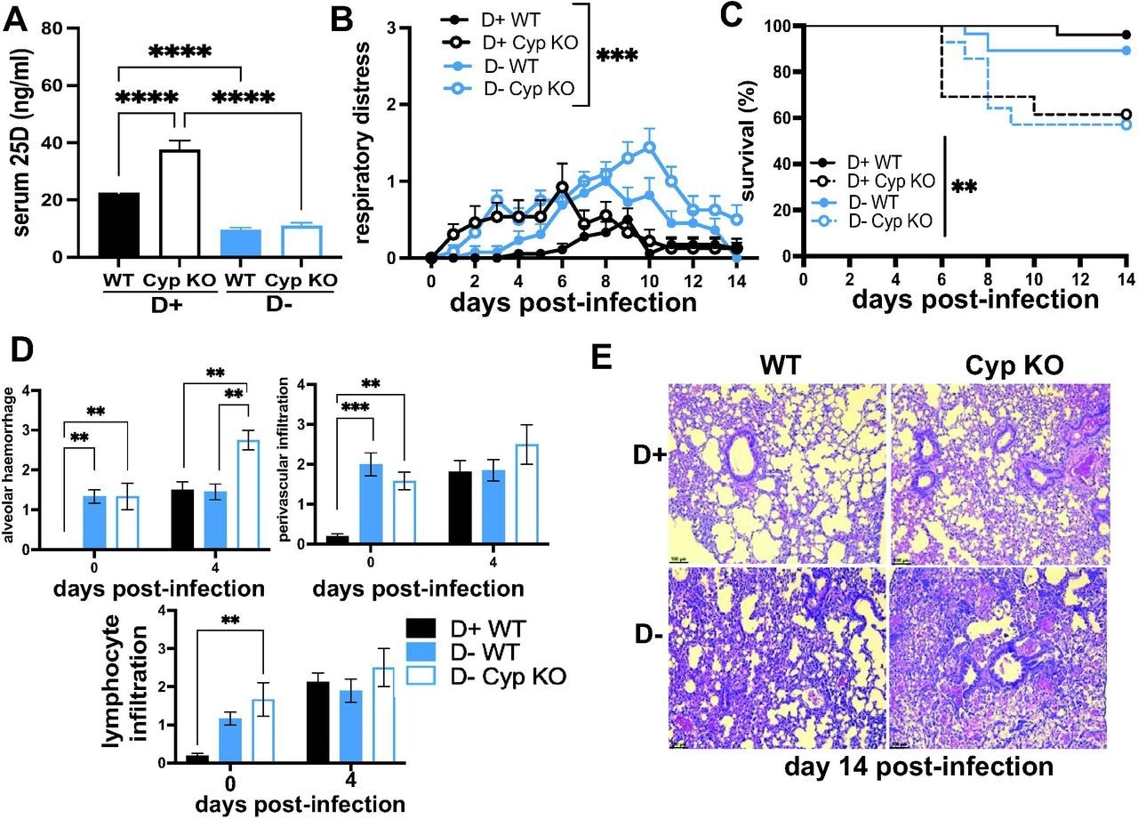
Vitamin D deficiency and Cyp27B1 KO increases susceptibility to H1N1 infection. D+ and D- WT and D+ and D- Cyp KO littermates were infected with H1N1 influenza (n=13-18 per group). A) Serum 25D, B)respiratory distress symptoms and C) survival of D+ WT (n=18), D+ Cyp KO (n=13), D- WT (n=13) and D- Cyp KO (n=12) mice at d14 post-infection. D) lung alveolar hemorrhage, perivascular infiltration and lymphocyte infiltration in D+ WT (n=4) and D- WT (n=4-5) and D- Cyp KO (n=2-3) mice at d0 and d4 post-infection. E) Representative histology of the lung of D+ WT (score = 3), D+ Cyp KO (score = 4), D- WT (score = 5.5) and D- Cyp KO (score = 6) at d14 post-infection. Values are the mean ± SEM. Statistical significance was assessed using one-way ANOVA with Bonferroni multiple comparison test for (A), two-way ANOVA with Bonferroni multiple comparison test for (B & D) and Log rank (Mankel-Cox) survival analysis for (C). **P < 0.01, ***P < 0.001 and ****P < 0.0001.
Vitamin D supplementation and SARS-CoV-2
SARS-CoV-2 infection and the effects of vitamin D supplementation were assessed in mice expressing the human angiotensin-converting enzyme 2 (ACE2) receptor, which is the receptor utilized by SARS-CoV-2 to gain entry into cells. Since Cyp27B1 remained present in this mouse strain, some mice were given a vitamin D deficient chow for eight weeks prior to infection to induce a state of vitamin D deficiency.
Vitamin D supplementation both before and after infection with SARS-CoV-2 led to significantly higher serum 25D levels as compared to vitamin D deficient mice. Meanwhile, vitamin D supplementation did not appear to impact survival rates following SARS-CoV-2 infection.
Mice consuming a vitamin D supplemented diet both before and after SARS-CoV-2 infection exhibited improved lung pathologies in terms of reduced expression of IFN-β, alveolar remodeling, type II hyperplasia, and total histopathology scores.
The researchers also evaluated the effects of vitamin D supplementation following SARS-CoV-2 infection in Golden Syrian hamsters. Similar to the mouse studies, the researchers also established a state of vitamin D deficiency in hamsters to better understand how vitamin D supplementation might improve SARS-CoV-2 infection outcomes in this environment.
To this end, serum-25D levels were comparable between hamsters fed on a chow diet (D+) and those fed on D- diets for four weeks. Furthermore, expression of the SARS-CoV-2 nucleocapsid (N) gene did not differ amongst D+ and D- lungs or colon.
Conclusions
A vitamin D deficient state appears to be vulnerable to viral infections due to the presence of pre-existing lung inflammation.
Taken together, the current study demonstrates that vitamin D supplementation reduces lung inflammation in mice infected with H1N1 and SARS-CoV-2. Notably, the beneficial effects of vitamin D supplementation in hamsters infected with SARS-CoV-2 were not remarkable. This lack of effect in hamsters may be due to the inability of commercially available chow to raise 25D levels.
Further studies are needed to describe the antiviral mechanism of vitamin D in preventing acute respiratory illnesses and the variations in effect if any, that its supplementation has in preventing H1N1 influenza and SARS-CoV-2 infection.
*Important notice
medRxiv publishes preliminary scientific reports that are not peer-reviewed and, therefore, should not be regarded as conclusive, guide clinical practice/health-related behavior, or treated as established information.
[if–>[if–>[if–>[if–>[if–>
[if–>[if–>[if–>[if–>[if–>[if–>[if–>[if–>[if–>[if–>[if–>[if–>[if–>[if–>[if–>[if–>[if–>[if–>
[if–>[if–>[if–>[if–>[if–>
[if–>[if–>[if–>[if–>[if–>[if–>[if–>[if–>[if–>[if–>[if–>[if–>[if–>[if–>[if–>[if–>[if–>[if–>
- Arora, J., Patel, D., Nicol, M., et coll. (2022). Vitamine D et capacité à produire du 1,25(OH)deuxD sont essentiels pour la protection contre l’infection virale des poumons. bioRxiv. doi:10.1101/2022.06.29.498158.